|
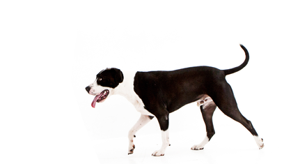
Gait analysis plays a vital role in canine rehabilitation, aiding veterinarians and canine rehabilitation specialists in understanding and treating various musculoskeletal conditions and injuries in dogs. By closely observing a dog's gait, experts can identify abnormalities and make informed decisions to develop effective treatment plans. In this blog post, we will explore the different forms of gait analysis in canine rehabilitation and look at the usability of each. 1. Visual Gait Assessment Visual gait assessment is the most common and basic form of gait analysis and is what I use the most. It involves observing a dog's movement visually to detect any abnormalities, asymmetries, or irregularities in their gait. I also tend to video all my dogs within the initial assessment so that I can go back later and slow the video down in case I have missed a small discrepancy. While this method is quick and easy to perform and requires little in the way of equipment, there is mixed evidence of the reliability of the data picked up by the therapist. A study by Evans and colleagues compared visual observation of gait to force plate analysis and evaluated 148 Labrador retrievers—131 that were 6 months post surgery for unilateral cranial cruciate ligament injury and 17 that were free of orthopaedic disease. The observer only identified 11% of the 131 dogs that were 6 months post surgery as being abnormal compared with force plate analysis, which revealed that 75% of the 131 dogs failed to achieve ground reaction forces consistent with sound Labrador retrievers. While force plate analysis is superior to visual observation, visual observation is still a practical tool in clinical practice, and its importance should not be discounted. 2. Pressure Mat Analysis; Kinetic movement
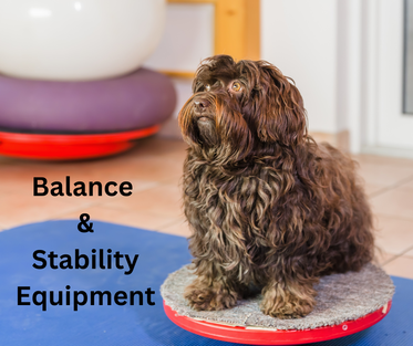
Pressure mat analysis involves the use of specialised mats equipped with sensors that measure the distribution of pressure and force exerted by a dog's paws during movement or at a stationary stance. This technique offers a quantitative assessment of gait parameters, providing detailed insights into weight distribution and symmetry. Pressure mat measurements have been the most widely used and validated quantitative gait application in veterinary medicine to date and are considered the 'gold standard' in gait analysis. However, there are disadvantages to force plate analysis. Limitations include the need for a long dedicated walkway, multiple trials, difficulty in setting up, breaking down, and moving, the complexity of software and data analysis and cost and impracticality for clinical practice. 3. Kinematic Gait Analysis Kinematic gait analysis involves utilising motion capture technology to record a dog's movements in three-dimensional space. Reflective markers placed on key anatomical landmarks allow for precise measurement of joint angles and movement patterns during gait. Even though this is a great way to measure the acceleration and velocity of body segments, it still has its disadvantages. This system is costly to set up along with user accuracy when placing the markers and there can be extra movement that is picked up of the markers on the dog's skin. Kinematic analysis also doesn't give reason to 'causes of movement dysfunction' unlike EMG as it only describes motion. 4. Electromyography (EMG) Electromyography involves measuring the electrical activity of muscles during gait. By analysing the timing and magnitude of muscle activation, researchers gain insights into muscle function and identify potential imbalances or weaknesses, particularly in the neurological dog. Gait analysis is a critical component of canine rehabilitation, enabling veterinarians and rehab therapists to evaluate the effectiveness of treatment plans and monitor a dog's progress. While visual gait assessment remains a fundamental tool, advancements in technology have introduced more sophisticated techniques like pressure mat analysis, kinematic gait analysis, and electromyography. Each approach contributes unique insights into a dog's gait, allowing for tailored rehabilitation strategies. Evans R, Horstman C, Conzemius M. Accuracy and optimization of force platform gait analysis in Labradors with cranial cruciate disease evaluated at a walking gait. Vet Surg 2005; 34(5):445-449. Gordon-Evans WJ. Gait analysis. In Tobias KM, Johnston SA (eds): Veterinary Surgery: Small Animal. St. Louis: Elsevier, 2012, pp 1190-1196. The bond between dogs and humans is growing stronger and more significant, leading to increased dedication by dog owners to provide the best possible care for their pets. Canine rehabilitation is becoming increasingly popular with dog owners as it provides a source of hope and healing for dogs dealing with various health conditions. In this blog post, we will examine some of the most commonly used tools in canine rehabilitation that I recommend. 1. Underwater Treadmills Underwater treadmills are one of the most effective tools in canine rehabilitation and are one of the reasons I chose to move away from a mobile business to a brick-and-mortar business. This technology allows dogs to exercise without bearing their full weight, reducing strain on their joints and muscles. The underwater treadmill therapy improves muscle strength and joint flexibility in dogs recovering from orthopaedic injuries and surgeries. I also love it as it is a functional movement and the buoyancy of water provides low-impact resistance, making it easier for canines to regain strength and mobility and is a firm favourite in my practice. 2. Therapeutic Ultrasound Therapeutic ultrasound is another modality that can be found in canine rehabilitation practice. This non-invasive treatment utilises high-frequency sound waves to promote tissue healing and helps with inflammation. I particularly use it on conditions such as ligament and tendon injuries or pathologies. 3. Balance and Stability Equipment Balance and stability equipment plays a vital role in enhancing a dog's core strength and proprioception. When it comes to balance equipment, there is a plethora to choose from! My go-to's are the inclined wedge, peanut stability ball and a dura disc. Stability balls and balance boards are great as they help dogs improve their balance and coordination, especially after neurological injuries. These tools challenge the dog's body to maintain stability, thereby strengthening muscles and improving overall stability. 4. Laser Therapy
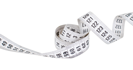
Laser therapy, also known as photobiomodulation or LLLT has gained popularity in canine rehabilitation due to its ability to alleviate pain and promote tissue repair. Laser therapy has also been shown to reduce pain and improve joint mobility in dogs with arthritis. The application of low-level laser light stimulates cellular activity, leading to accelerated healing and reduced inflammation. Out of any modality in my clinic, the laser is my most favourite. It is non-invasive and non-painful to administer. It also doesn't require a gel (unlike the ultrasound machine) to use it. 5. Therapeutic Massage Therapeutic massage has long been recognised as a beneficial treatment for both humans and animals. Massage therapy can reduce muscle tension and improve the flexibility of tissues, particularly with dogs that have musculoskeletal conditions. Skilled massage techniques help increase circulation, reduce scar tissue, and enhance the release of endorphins, all of which contribute to the dog's overall well-being. 6. Assisted Rehabilitation Devices Assisted rehabilitation devices, such as harnesses and slings, are instrumental in supporting dogs during the recovery process. There are several ones to choose from however the one I reach for the most would have to be the help 'em up harness. This harness is adjustable in all aspects, has a front and rear fit (which can be detached if needed) and has strong steady handles to help assist any dog in need of a helping hand. I find that in practice, dogs that need the help em up harnesses regain their confidence and function faster than those that don't. The proper use of harnesses and slings assists dogs in walking and exercising, preventing further injury while building strength. The field of canine rehabilitation has made great strides in improving the quality of life for dogs. The most effective tools that I use in practice include underwater treadmills, therapeutic ultrasound, balance and stability equipment, laser therapy, therapeutic massage, and assisted rehabilitation devices. By utilising the latest tools and techniques in canine rehabilitation, rehab therapists can help dogs lead a healthier and happier life. Staying informed about the latest research and advancements in this field is essential to providing the best care possible for our dogs, enriching their lives and ours A dog's muscle mass plays a significant role in its health and physical abilities. To ensure proper monitoring of strength and detect any muscular abnormalities, objective assessment of muscle mass is crucial, particularly if you are a physiotherapist or rehab therapist. While various methods are available, the most prevalent way to measure muscle mass in dogs is through the thigh circumference using a tape measure. Physiotherapists and rehabilitation therapists often take note of this measurement to establish a baseline before implementing a strength and conditioning program. They will then re-evaluate the measurement at a later date to determine if there are satisfactory improvements in muscle mass and strength. How reliable is this measurement and what factors can influence the readings between people and between repeat measurements?References:
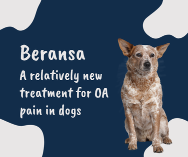
Bascuñán, A. L., Kieves, N., Goh, C., Hart, J., Regier, P., Rao, S., Foster, S., Palmer, R., & Duerr, F. M. (2016, May 5). Evaluation of Factors Influencing Thigh Circumference Measurement in Dogs. Journal Name, 1st published, 2016-05-05 Smith, T. J., Baltzer, W. I., Jelinski, S. E., & Salinardi, B. J. (2015). Inter- and Intratester Reliability of Anthropometric Assessment of Limb Circumference in Labrador Retrievers. Journal Name, 1st published, 08 October 2015 Arthritis affects millions of dogs worldwide and is something that I see, as a physiotherapist, on the regular in practice. Symptoms range from slow to rise, avoidance of certain tasks such as jumping up on the couch, changes in behaviour, lameness and many more. The continuous search for effective treatment options has led to the development of Beransa (known as Librela overseas) which has finally made it's way to Australia shores and is currently being used to treat pain in arthritic dogs. So what exactly is Beransa? Beransa, delivered as a once monthly subcutaneous injection, delivers a big boost of monoclonal antibodies - a type of immune system protein. In a simplistic way, the monoclonal antibodies go on to mimic naturally occurring antibodies and begin to block pain signals due to it's unique ability to attach to nerve growth factor. Nerve growth factor is an essential protein which is important for growth and survival of sensory nerves (which needs to attach to it's receptor on a nerve cells to transmit pain signals) and voila, just like that the nerve cannot attach to nerve growth factor to transmit it's pain signal because the monoclonal antibody has decided to take up residency and couple with nerve growth factor instead. When did Beransa become available in Australia?
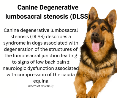
Beransa was first made available in Australia this year (2023) (and in Europe in 2021) and is a fairly new addition to the treatment of OA pain in dogs. Since its introduction, veterinarians and pet owners have been in favour of it due to it's reported low adverse reaction rate. What is the cost of administration? The cost of administering Beransa can vary depending on the dog's size, the severity of arthritis, and the chosen treatment plan. As with any specialised medication, dog owners should consult their veterinarian to determine the most suitable dosage and duration of treatment for their pet. Beransa can be a tad more expensive compared to some common traditional treatments. Are there any potential side effects? Like any medication, Beransa may have some potential adverse effects on dogs. However, research has shown that these adverse effects are generally mild and well-tolerated. The most common adverse events reported include urinary tract infections, bacterial skin infections, dermatitis, and increased blood urea nitrogen. Beransa should also not be administered to breeding, pregnant or lactating dogs. What is the research saying? In two field studies, dogs administered Beransa as a monthly injection demonstrated a reduction in OA pain compared to dogs that received the placebo and by reducing pain, Beransa was also shown to help their mobility and overall quality of life (corral et al 2022) While effectiveness may not be seen until after the second dose of Beransa, some dogs may experience a reduction in pain as early as seven days after the first dose. Additionally, in a continuation study, dogs treated with Beransa experienced lasting OA pain relief over the course of the study with monthly injections (Corral et al 2022) European veterinarians who have used Beransa rated their overall satisfaction 8.6 out of 10, the highest of any OA pain medication evaluated (ZMR 2022) Beransa, represents a promising breakthrough in the treatment of canine arthritis and offers hope for significantly improving the comfort and well-being of arthritic dogs by reducing pain associated with arthritis. With it's relatively low reported adverse reaction/ side effects and overall high satisfaction rates by veterinarians, Beransa is fast becoming a favourite in treating OA pain in dogs. References: Corral, M. J., et al. (2021). A prospective, randomized, blinded, placebo-controlled multisite clinical study of bedinvetmab, a canine monoclonal antibody targeting nerve growth factor, in dogs with osteoarthritis. Veterinary Anaesthesia and Analgesia, 48, 943-955. U.S. Food and Drug Administration (FDA). (2023, May 5). FDA Approves First Monoclonal Antibody for Dogs with Osteoarthritis Pain. Retrieved May 13, 2023. DLSS is a condition that affects the spinal canal, leading to the narrowing of the space and compression of the spinal cord and nerves. Dogs can suffer from this debilitating condition, which can cause pain, weakness, and a decline in their quality of life. In this blog post, we will delve into the causes, symptoms, and treatment options for spinal stenosis in dogs, shedding light on the importance of early detection and specialised care. Understanding Spinal Stenosis: Canine degenerative lumbosacral stenosis (DLSS) describes a syndrome in dogs associated with degeneration of the structures of the lumbosacral junction leading to signs of low back pain ± neurologic dysfunction associated with compression of the cauda equina. DLSS has a multifactorial origin in which intervertebral disc (IVD) degeneration plays a major role. However, the DLSS syndrome lacks pathognomonic characteristics, and diagnosis is often presumptive based on a combination of clinical signs, findings on advanced imaging, and ruling out other specific etiologies that cause cauda equina compression (Worth et al 2019) Incidence and prevalence of spinal stenosis in dogs: While exact figures for the incidence and prevalence of spinal stenosis in dogs are challenging to determine, research suggests that certain breeds are more susceptible. In general breeds like German Shepherds, and Doberman Pinschers are at a higher risk due to their anatomical conformation and genetic factors Recognising the symptoms:
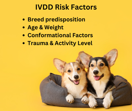
.Detecting spinal stenosis in its early stages is crucial for successful management. Dogs with spinal stenosis may exhibit symptoms such as back pain, difficulty with certain movements, loss of coordination, hind limb weakness, and, in severe cases, paralysis. Prompt recognition of these signs can help initiate appropriate treatment and improve outcomes. Diagnostic Techniques: Accurate diagnosis is key to developing an effective treatment plan. Veterinary professionals may employ diagnostic imaging techniques such as radiography, computed tomography (CT), or magnetic resonance imaging (MRI) to evaluate the extent of stenosis, identify underlying causes, and assess the impact on the spinal cord and nerves Treatment Approaches: The treatment of spinal stenosis in dogs aims to alleviate pain, improve mobility, and enhance the overall quality of life. The specific treatment approach depends on the type and severity of stenosis. Treatment options may include: a. Medication: Pain management medication, such as non-steroidal anti-inflammatory drugs (NSAIDs) or analgesics, may be prescribed to reduce inflammation and alleviate discomfort. b. Physiotherapy and Rehabilitation: Canine rehabilitation therapists play a vital role in managing spinal stenosis. Through tailored treatment plans, they focus on pain management, range of motion exercises, hydrotherapy, and strengthening activities to enhance the dog's mobility and promote healing. c. Surgery: In more severe cases of spinal stenosis, surgical intervention may be necessary. Surgical options include decompression, distraction stabilisation technique or laminectomy, which involves removing the bone or tissue that is compressing the spinal cord, or spinal fusion to stabilise the affected area. Home care and long term management: Continued care at home is essential for the long-term management of spinal stenosis. Canine physiotherapists educate owners on exercises, environmental modifications, and assistive devices that can support the dog's mobility and comfort. Compliance with prescribed home care is crucial for maximising the benefits of rehabilitation therapy Spinal stenosis in dogs can significantly impact their mobility, comfort, and overall quality of life. Canine physiotherapists play a vital role in managing this condition, offering non-surgical alternative Spinal stenosis can significantly impact a dog's mobility and overall well-being. Early detection, accurate diagnosis, and a comprehensive treatment approach are key to managing this condition effectively. By working closely with veterinary professionals and canine physiotherapists, owners can provide their dogs with the specialised care needed to alleviate pain, improve mobility, and enhance their quality of life. References: Worth et al (2019) Canine Degenerative Lumbosacral Stenosis: Prevalence, Impact And Management Strategies Canine Intervertebral Disc Disease (IVDD) is a prevalent and potentially debilitating condition that affects the spinal discs of dogs. IVDD occurs when the intervertebral discs, which act as cushions between the vertebrae, degenerate or become herniated. This condition can cause pain, mobility issues, and, in severe cases, paralysis. In this blog post, we will delve into the causes, risk factors, and preventative measures associated with IVDD in dogs To grasp the implications of IVDD, it is crucial to comprehend the structure and function of intervertebral discs. These discs have a tough outer layer called the annulus fibrosus and a soft, gelatinous center known as the nucleus pulposus. IVDD can manifest in two primary forms: a. Hansen Type I: This acute form is characterized by a sudden rupture or protrusion of the disc material into the spinal canal, leading to compression of the spinal cord. b. Hansen Type II: This chronic form involves gradual degeneration of the disc, leading to herniation or bulging over time. There are several factors that increase the risk of Intervertebral Disc Disease (IVDD) in dogs. These factors are as follows:
Breed Predisposition: Certain dog breeds have a higher susceptibility to IVDD due to genetic factors. Dachshunds, Shih Tzus, Beagles, and Pekingese are among the breeds that are at an increased risk, according to a study published in the Journal of Veterinary Internal Medicine (Kealy et al., 2012). Age and Weight: Older dogs and overweight or obese individuals are more likely to develop IVDD. This suggests that age and weight are significant risk factors. Conformational Factors: Dogs with certain physical attributes, such as long backs and short legs, are more likely to get IVDD. Breeds like Basset Hounds and French Bulldogs are examples of this conformational predisposition. Trauma and Activity Level: Traumatic incidents like falls or excessive jumping increase the risk of disc damage. Additionally, high-impact activities that strain the spine, like repetitive jumping or twisting, can worsen the condition. Preventive Measures: Canine Intervertebral Disc Disease (IVDD) is a common condition that can affect a dog's quality of life. While not all cases of IVDD can be prevented, there are measures that pet owners can take to reduce the risk or severity of this condition. These include: Weight management: Keeping a healthy weight through proper nutrition and regular exercise can help reduce stress on the intervertebral discs. Controlled exercise: Encouraging controlled exercise and avoiding high-impact activities can help minimize the strain on the spine. Regular walks, swimming, and low-impact play are beneficial for dogs at risk of IVDD. Environmental modifications: Providing supportive bedding, avoiding high surfaces, and using ramps or stairs can reduce the risk of falls or jumping-related injuries. Regular veterinary check-ups: Routine examinations allow veterinarians to monitor the dog's spinal health and detect early signs of IVDD, enabling prompt intervention. As always, if you are reading this blog post as a pet owner, always consult with a veterinarian or physiotherapist for tailored advice based on your dog's individual needs and breed predisposition. By understanding the risk factors associated with IVDD and implementing preventive strategies, we can help safeguard the spinal health and overall well-being of our doggos. References: Kealy, R. D., Lawler, D. F., Ballam, J. M., Mantz, S. L., Biery, D. N., Greeley, E. H., Lust, G., Segre, M., Smith, G. K., & Stowe, H. D. (2012). Effects of diet restriction on life span and age-related changes in dogs. Journal of the American Veterinary Medical Association, 220(9), 1315–1320. Courcier, E. A., Thomson, R. M., Mellor, D. J., & Yam, P. S. (2010). An epidemiological study of environmental factors associated with canine obesity. Journal of Small Animal Practice, 51(7), 362–367. |
AuthorJoanna Whitehead Archives
June 2024
Categories
All
|

 RSS Feed
