
|
Canine hip dysplasia (CHD) is a common orthopaedic condition affecting dogs, particularly large and giant breeds. It is a multifactorial disorder characterised by abnormal development and/or degeneration of the hip joint. In this blog post, we will delve into the details of canine hip dysplasia and explore the risk factors associated with it. 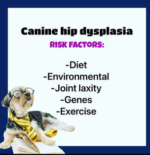 Canine hip dysplasia risk factors Canine hip dysplasia occurs when the hip joint fails to develop properly. This leads to a loose and unstable joint, causing abnormal wear and tear, inflammation, and eventually, degenerative joint disease. Over time, the condition can result in pain, lameness, and reduced mobility for affected dogs. Risk Factors for Canine Hip Dysplasia: 1) Genetic Predisposition: Genetic factors play a significant role in the development of hip dysplasia. Certain breeds have a higher incidence of CHD, including Labrador Retrievers, German Shepherds, Golden Retrievers, Rottweilers, and Great Danes. These breeds often have a genetic predisposition to the condition, making it more likely to be passed on to their offspring. According to a study by Smith et al. (2019), "Prevalence of canine hip dysplasia in purebred dogs in North America," certain breeds exhibited a higher prevalence of CHD, further highlighting the genetic influence. 2) Growth Rate and Nutrition: Rapid growth during puppyhood can contribute to the development of hip dysplasia. Overfeeding or an imbalanced diet that lacks essential nutrients can accelerate the growth rate, putting additional stress on the developing hip joint. Excessive weight gain can exacerbate the condition. A study conducted by Kealy et al. (1997) titled "Effects of limited food consumption on the incidence of hip dysplasia in growing dogs" found that restricted feeding resulted in a decreased incidence of CHD in Labrador Retrievers. 3) Obesity: Obesity is a major risk factor for hip dysplasia. The extra weight places increased strain on the hip joint, leading to accelerated degeneration and worsening of the condition. Maintaining a healthy body weight can help reduce the risk and alleviate symptoms. A study published by Kasström et al. (2014) titled "Effects of conformational and environmental risk factors on hip dysplasia in German Shepherd dogs in Sweden" identified obesity as a significant risk factor for CHD. 4) Environmental Factors: Environmental factors, such as excessive exercise on hard surfaces, can contribute to the development of CHD. Activities that involve repetitive impact and high-intensity exercise at a young age may negatively impact hip joint development. A study by Smith et al. (2018) titled "Canine hip dysplasia: Understanding and applying epidemiologic evidence to improve canine welfare" highlights the influence of environmental factors on the occurrence of CHD. Prevention and Management: While canine hip dysplasia has a strong genetic component, certain measures can help reduce the risk and manage the condition effectively. Responsible breeding practices, including hip screening programs, can help identify and reduce the occurrence of CHD in susceptible breeds. Additionally, providing balanced nutrition, maintaining a healthy weight, and avoiding excessive high-impact exercise during the critical growth period can aid in prevention. Canine hip dysplasia is a prevalent condition that affects many dogs, particularly those of larger breeds. Understanding the risk factors associated with CHD is crucial for both breeders and dog owners. By identifying these factors and implementing preventive measures, such as responsible breeding and appropriate nutrition, we can mitigate the impact of hip dysplasia. References:
Elbow dysplasia is a common orthopaedic condition that affects dogs, particularly large and giant breeds. It refers to a group of developmental abnormalities that occur in the elbow joint, leading to pain, lameness, and reduced mobility. In this blog post, we will explore the different types of canine elbow dysplasia, their symptoms, and the available treatment options. 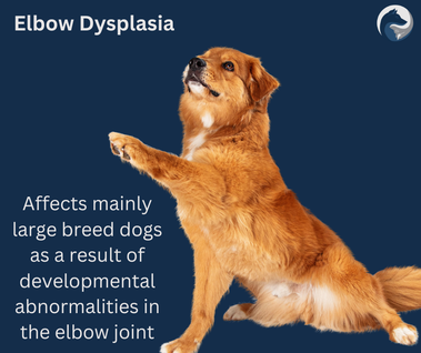
Canine elbow dysplasia encompasses various conditions that can affect a dog's elbow joint, causing pain and reduced mobility. Early detection, accurate diagnosis, and appropriate treatment are crucial for improving the quality of life for affected dogs. With the right approach, many dogs can lead happy, active lives. Patella luxation, also, is a common orthopaedic condition in dogs. In fact out of all the doggos I see, the top of the list are those with MPL (medial patella luxation) or post surgery from this. Several risk factors can contribute to the development of patella luxation in dogs. Here are some of the main risk factors: 1. Breed Predisposition: Certain dog breeds are more prone to patella luxation. Small and toy breeds such as Chihuahuas, Pomeranians, Pekingese, Yorkshire Terriers, and Miniature Poodles have a higher incidence of this condition. Some larger breeds like Labrador Retrievers and Akitas can also be affected. 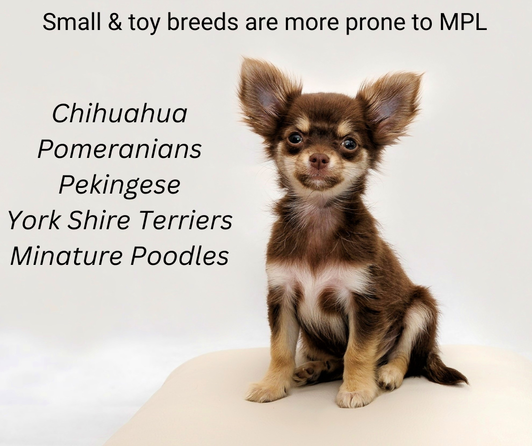 Small and toy breeds are more prone to MPL 2. Genetic Factors: Patella luxation can have a genetic component, meaning it can be passed down from parent dogs to their offspring. Breeding dogs with a history of patella luxation increases the likelihood of the condition in their offspring.
3. Congenital Abnormalities: Dogs born with abnormal bone structure or joint development are at a higher risk of patella luxation. Factors such as shallow femoral grooves, misaligned bones, and abnormal patellar ligaments can contribute to the condition or having bowed legs. 4. Obesity: Overweight or obese dogs are more prone to patella luxation due to the added stress on their joints. Excess weight puts strain on the patellar ligaments and can increase the likelihood of the kneecap slipping out of place. 5. Trauma: Injuries or trauma to the knee area can cause patella luxation in dogs. Traumatic incidents such as accidents, falls, or rough play can result in dislocation of the kneecap. 6. Muscle Weakness: Weak thigh muscles or muscles around the stifle joint can contribute to patella luxation. When the muscles are not adequately developed or are imbalanced, they may fail to provide proper support and stability to the patella. 7. Age: Patella luxation can occur at any age, but it is more commonly seen in young dogs, especially those under one year of age. In some cases, the condition may worsen as the dog ages. It's important to note that while these risk factors increase the likelihood of patella luxation, they do not guarantee that a dog will develop the condition. Regular veterinary check-ups, maintaining a healthy weight, and providing appropriate exercise can help reduce the risk of patella luxation in dogs. Canine cruciate disease, also known as cranial cruciate ligament (CCL) disease, is a common orthopaedic condition that affects dogs. The cruciate ligament is a crucial stabilising structure within the stifle (knee) joint, and when it becomes damaged or ruptured, it leads to instability and lameness. 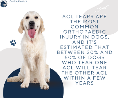 Incidence of CCL (ACL) tears in dogs The exact cause of canine cruciate disease is not fully understood, but it is believed to be a combination of genetic predisposition and various risk factors. Unlike human ACL's that tend to acutely rupture, a dogs cruciate tends to be more progressive and degenerative in nature Here are some of the main factors that can contribute to the development of canine cruciate disease:
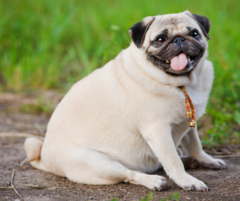 Obesity in dogs can be a risk factor in CCL disease 4. Conformation: Certain anatomical features and conformational abnormalities can increase the risk of cruciate disease. Dogs with a steep tibial plateau angle, shallow knee joint, or other structural abnormalities such as 'upright hind limbs' or 'bowed legs' can be more susceptible to CCL disease.
5. Trauma: Acute injury or trauma to the knee joint, such as jumping, landing awkwardly, or sudden twisting motions, can cause cruciate ligament damage. Traumatic events may contribute to the development of cruciate disease in some cases. 6. Hormonal factors: There is some evidence to suggest that female dogs that have not been spayed may have an increased risk of developing cruciate disease. The exact relationship between hormones and ligament health is not fully understood. It's important to note that while these factors can increase the likelihood of developing cruciate disease, it can still occur in dogs without any apparent risk factors. If you happen to suspect that your dog may have CCL disease, please take them to your friendly vet for an accurate diagnosis. |
AuthorJoanna Whitehead Archives
June 2024
Categories
All
|
 RSS Feed