
|
Canine cranial cruciate ligament (CCL) injuries are a commonly occurring condition among dogs, which can cause them to experience discomfort, reduced mobility, and lameness. Tibial Plateau Leveling Osteotomy (TPLO) surgery is a highly effective surgical treatment available for this condition. TPLO surgery aims to stabilise the knee joint and restore function, enabling your doggo friend to return to an active, pain-free lifestyle. However, the success of TPLO surgery greatly depends on the efforts of prehabilitation (prehab) and rehabilitation (rehab). In this blog, we will delve into what TPLO surgery entails and explore the prehab and rehab options to ensure a smooth and successful recovery journey for your beloved doggo companion. TPLO surgery is a surgical procedure that aims to restore stability and function to the knee joint of dogs with CCL injuries. The surgery involves reshaping the tibial plateau, which helps the joint regain balance and stability. This surgery provides a new hope for dogs struggling with pain, discomfort, and mobility issues. Before the surgery, the dog undergoes a careful assessment to determine the best surgical approach. The surgery is performed under anaesthesia, and the surgeon makes an incision to reach the knee joint. The surgeon then carefully reshapes the tibial plateau, using specialised implants to ensure stability and protect the joint. After the surgery, the dog requires rehabilitation to regain mobility and strength. The veterinary team will design a rehabilitation program tailored to the dog's unique needs, which may include exercises, manual therapy, and other modalities. Postoperative challenges such as infection, implant failure, and delayed healing may occur, but the veterinary team is always ready to address these challenges and guide the dog towards recovery. 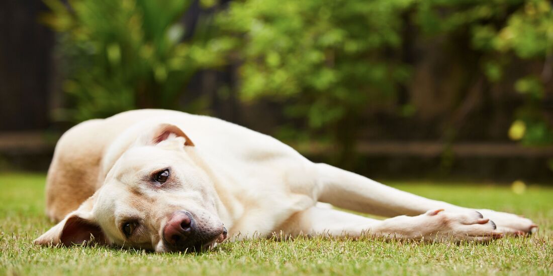 Prehabilitation Prehabilitation, also known as prehab, plays a crucial role in optimising your dog's physical condition and mental readiness before undergoing Tibial Plateau Leveling Osteotomy (TPLO) surgery. By implementing a comprehensive prehabilitation program, you can enhance your dog's overall well-being, improve surgical outcomes, and facilitate a smoother recovery process. Consultation with a Veterinary Professional - Schedule a consultation with your veterinarian or a certified veterinary surgeon to discuss your dog's condition, treatment options, and prehabilitation plan. - Seek guidance on preoperative requirements, including diagnostic tests, blood work, and any necessary medications or supplements. Weight Management - Evaluate your dog's body condition score and work with your veterinarian to establish a target weight range. - Implement a tailored diet plan to achieve gradual weight loss if necessary, focusing on portion control, high-quality nutrition, and regular exercise. - Monitor your dog's weight regularly and adjust the diet plan as needed to maintain progress towards the target weight. Muscle Strengthening - Incorporate targeted exercises to strengthen the muscles surrounding the affected knee joint, including the quadriceps, hamstrings, and hip abductors. - Engage in low-impact activities such as leash walks, controlled stair climbing, and swimming to promote muscle development and joint stability. - Consult with a certified canine rehabilitation therapist for personalized exercise recommendations and guidance on proper technique. Joint Supplements - Discuss the use of joint supplements with your veterinarian to support your dog's joint health and mobility. - Consider incorporating supplements such as glucosamine, chondroitin, omega-3 fatty acids, and antioxidants into your dog's daily regimen. - Choose high-quality supplements specifically formulated for canine use and follow the recommended dosage instructions provided by the manufacturer. Mental Preparation - Introduce your dog to handling procedures, such as gentle manipulation of the affected leg, to familiarise them with prehabilitation exercises and minimise stress on the day of surgery. - Utilise positive reinforcement training techniques to create a positive association with prehabilitation activities and promote cooperation and relaxation. - Provide environmental enrichment, interactive toys, and mental stimulation to engage your dog and reduce anxiety during prehabilitation. Environmental Modification - Create a safe and comfortable environment for your dog to rest and recuperate before surgery. - Minimise environmental stressors, such as loud noises or unfamiliar visitors, to promote relaxation and reduce anxiety. - Ensure easy access to food, water, and a comfortable resting area to support your dog's physical and emotional well-being. Veterinary Follow-Up - Schedule regular follow-up appointments with your veterinarian to monitor your dog's progress and adjust the prehabilitation plan as needed. - Immediately communicate any changes in your dog's condition, behaviour, or response to prehabilitation activities to your veterinary team. - Collaborate closely with your veterinarian and veterinary surgeon to ensure your dog receives comprehensive care and support throughout the prehabilitation process. By following this prehabilitation guide and working closely with your veterinary team, you can empower your canine companion to face TPLO surgery with confidence and resilience. By optimising your dog's physical condition, mental well-being, and overall readiness for surgery, you can enhance the likelihood of a successful outcome and promote a smoother recovery journey. 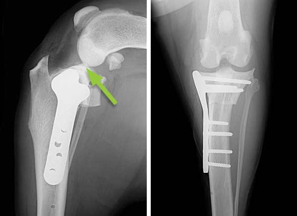 Rehabilitation Rehabilitation following Tibial Plateau Leveling Osteotomy (TPLO) surgery is essential for promoting healing, restoring mobility, and ensuring the long-term success of the procedure. By implementing a structured rehabilitation program, you can support your dog's recovery journey, minimise complications, and optimise their return to an active, pain-free lifestyle. Consultation with a Certified Canine Rehabilitation Therapist - Schedule a consultation with a certified canine rehabilitation therapist (CCRT) or a veterinary professional experienced in postoperative care. - Discuss your dog's surgical procedure, postoperative restrictions, and rehabilitation goals to develop a tailored rehabilitation plan. Pain Management - Administer prescribed pain medications as directed by your veterinarian to manage postoperative discomfort and inflammation. - Monitor your dog's pain levels closely and communicate any changes or concerns to your veterinary team promptly. - Explore additional pain management modalities, such as cold therapy, laser therapy, or acupuncture, under the guidance of a qualified professional. Controlled Exercise - Begin a structured exercise program focusing on controlled movement, muscle strengthening, and joint flexibility. - Gradually increase the duration and intensity of exercise sessions while monitoring your dog's response and adjusting the program as needed. - Incorporate low-impact activities such as leash walking, hydrotherapy, and therapeutic exercises to promote joint mobility and muscle recovery. Rehabilitation Exercises - Engage in targeted rehabilitation exercises designed to improve proprioception, balance, and joint stability. - Include exercises such as sit-to-stand transitions, weight shifting, balance exercises on unstable surfaces, and gentle range of motion exercises. - Work closely with your rehabilitation therapist to ensure proper technique and progression of exercises based on your dog's individual needs and capabilities. Joint Protection - Implement strategies to protect the surgical site and minimize stress on the affected limb during the rehabilitation process. - Use supportive devices such as orthopaedic braces, bandages, or protective boots as recommended by your veterinary team. - Avoid activities that place excessive strain on the surgical site, such as running, jumping, or rough play, until cleared by your veterinarian. Nutritional Support - Provide a balanced diet rich in high-quality protein, vitamins, and minerals to support your dog's recovery and tissue healing. - Consider incorporating joint supplements such as glucosamine, chondroitin, omega-3 fatty acids, and antioxidants to promote joint health and reduce inflammation. - Consult with your veterinarian to ensure that your dog's nutritional needs are met during the rehabilitation period. Monitoring and Progress Evaluation - Monitor your dog's progress closely, noting improvements in mobility, muscle strength, and comfort levels. - Attend follow-up appointments with your veterinary team to assess your dog's healing progress, address any concerns, and adjust the rehabilitation plan as needed. - Keep a journal or log of your dog's rehabilitation activities, pain levels, and milestones achieved to track progress and inform future decisions. Patience and Support - Be patient and supportive as your dog navigates the challenges of rehabilitation and recovery. - Offer encouragement, praise, and rewards for participating in rehabilitation exercises and maintaining a positive attitude. - Provide a calm and nurturing environment free from stressors to promote relaxation and well-being during the recovery process. By following this rehabilitation guide and working closely with your veterinary and rehabilitation team, you can facilitate a successful recovery for your doggo companion following TPLO surgery. With dedication, patience, and proper support, you can help your dog regain mobility, strength, and vitality, paving the way for a happy and active life post-surgery.  TPLO surgery offers a promising solution for canine CCL injuries, but successful outcomes depend on diligent prehabilitation and rehabilitation efforts. You can optimise their recovery and enhance their quality of life by implementing appropriate prehab strategies to prepare your dog for surgery and following a structured rehab program postoperatively. Consult with your veterinarian or a certified canine rehabilitation therapist to develop a customised prehab and rehab plan tailored to your dog's individual needs. With proper care and support, your doggo friend can return from TPLO surgery and enjoy an active, pain-free lifestyle again.
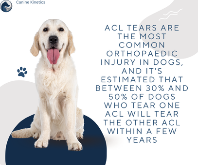
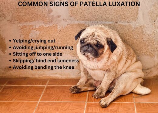
If you want to learn more tricks and tips and be involved in a community of like-minded individuals, join our Facebook group at: Canine knee injuries are a common concern among dog owners, affecting various breeds and sizes. These injuries can significantly impact a dog's mobility, comfort, and quality of life. Understanding the different types of knee injuries and the pathways for rehabilitation is crucial for ensuring the best possible outcomes for our doggo companions. Types of Canine Knee Injuries Cruciate Ligament Tears The cruciate ligaments, namely the cranial cruciate ligament (CCL), are vital for stabilising the knee joint in dogs. Tears or ruptures in the CCL are among the most common knee injuries in canines, often resulting from sudden trauma or degenerative changes. Dogs with cruciate ligament tears typically exhibit lameness, swelling, and difficulty bearing weight on the affected limb. Patellar Luxation Patellar luxation involves the dislocation of the kneecap (patella) from its normal position within the groove of the femur. This condition can be congenital or acquired and may vary in severity from intermittent to permanent dislocation. Symptoms include intermittent limping, skipping, or "locking" of the knee joint. Meniscal Injuries The menisci are cartilage structures within the knee joint that provide cushioning and stability. Injuries to the menisci commonly accompany cruciate ligament tears but can also occur independently. Dogs with meniscal injuries may exhibit lameness, joint swelling, and pain, particularly during movement. Rehabilitation Pathways Conservative Management Conservative management is an approach to treating canine knee injuries without resorting to surgical intervention. This method is often considered in cases where surgery may not be feasible or necessary, such as in older dogs, those with concurrent health issues that increase surgical risks, or in instances where the injury is mild to moderate. Here's a more in depth exploration of conservative management strategies for canine knee injuries.
Weight Management Maintaining a healthy body weight is crucial for dogs with knee injuries, as excess weight can exacerbate strain on the joint and impede healing. A balanced diet, portion control, and appropriate exercise regimens tailored to the dog's needs can help achieve and maintain a healthy body weight. Pain Management Pain relief is an integral component of conservative management and may involve the use of non-steroidal anti-inflammatory drugs (NSAIDs) prescribed by a veterinarian to alleviate discomfort and reduce inflammation. Other pain management modalities, such as joint supplements (e.g., glucosamine and chondroitin), nutraceuticals, or alternative therapies like acupuncture, may also be considered to enhance pain relief and support joint health. Physiotherapy and Rehabilitation Physiotherapy modalities can play a significant role in conservative management by promoting healing, reducing pain, and improving joint function. Common physiotherapy techniques include:
Orthopaedic Supports Orthopaedic devices, such as knee braces or supportive wraps, may be recommended in some cases to provide additional stability and support to the injured knee joint. These supports can help alleviate discomfort, reduce inflammation, and improve proprioception while the knee heals. Regular Monitoring and Follow-Up Close monitoring of the dog's progress is essential during conservative management, with regular follow-up appointments scheduled to assess response to treatment and make any necessary adjustments to the management plan. Radiographic imaging or other diagnostic tests may be repeated periodically to evaluate healing and monitor for any changes in the condition of the knee joint. Conservative management can be an effective approach for managing certain canine knee injuries, particularly in cases where surgery may not be indicated or desired. However, it's important to work closely with a veterinarian and rehabilitation expert to develop a comprehensive treatment plan tailored to the individual dog's needs and condition. With proper care, many dogs can experience significant improvement in their mobility and quality of life through conservative management strategies. 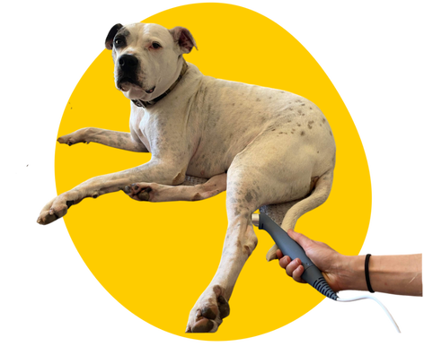 Surgical Intervention
Surgical intervention is often recommended for severe or progressive knee injuries, especially cruciate ligament tears and advanced cases of patellar luxation. Common surgical procedures include:
Tibial Plateau Leveling Osteotomy (TPLO) TPLO is a surgical procedure primarily used to address cranial cruciate ligament (CCL) tears, which are one of the most common knee injuries in dogs. During TPLO, the veterinarian makes an incision over the knee joint and cuts the top portion of the tibia (shinbone). The angle of the tibial plateau is then altered and stabilised using specialised plates and screws, effectively changing the biomechanics of the knee joint. By altering the angle of the tibial plateau, TPLO reduces the need for the cranial cruciate ligament, providing stability to the knee joint during weight-bearing activities. TPLO is known for promoting faster and more reliable healing compared to some other surgical techniques. Cranial Cruciate Ligament Repair (CCLR) CCLR involves repairing or replacing the torn cranial cruciate ligament (CCL) to restore stability to the knee joint. There are several techniques used for CCLR, including:
Patellar Realignment Procedures Patellar luxation, where the kneecap (patella) dislocates from its normal position, may require surgical intervention if conservative measures fail to provide relief. Surgical procedures for patellar luxation aim to realign the patella and correct any underlying anatomical abnormalities contributing to the dislocation. Common procedures include:
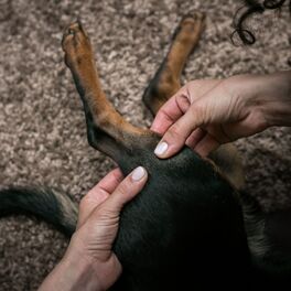
The specific procedure chosen depends on factors such as the severity of the luxation, concurrent orthopaedic issues, and the dog's size and breed. These surgical procedures aim to restore stability and function to the knee joint, alleviate pain, and improve the dog's overall quality of life. It's essential to discuss the available surgical options with a veterinarian or veterinary surgeon to determine the most suitable approach based on the individual needs of the dog. Additionally, post-operative rehabilitation is crucial for optimising recovery and maximising the success of the surgical intervention. Post-Surgical Rehabilitation Post-operative rehabilitation plays a vital role in the recovery process following surgical intervention for canine knee injuries. Below is and extended and detailed exploration of rehabilitation strategies and modalities. Controlled Exercise Regimens Controlled exercise is crucial during the rehabilitation period to promote healing, rebuild strength, and restore normal joint function. Initially, exercise should be limited to short, leash-controlled walks to prevent excessive strain on the surgical site. As the dog progresses, the veterinarian or rehabilitation specialist may recommend gradually increasing the duration and intensity of exercise, incorporating activities such as controlled leash walking, gentle jogging, and low-impact exercises. It's essential to avoid activities that may put undue stress on the knee joint, such as jumping, running on slippery surfaces, or rough play, especially during the early stages of rehabilitation. Joint Mobilisation and Range of Motion Exercises Joint mobilisation techniques, including passive range of motion exercises, help maintain or improve joint flexibility and prevent stiffness. These exercises may involve gently moving the affected joint through its full range of motion, guided by a trained rehabilitation therapist. Range of motion exercises should be performed regularly but carefully to avoid overstretching or causing discomfort to the dog. Therapeutic Modalities Various therapeutic modalities can aid in pain management, reduce inflammation, and promote tissue healing. Common modalities include:
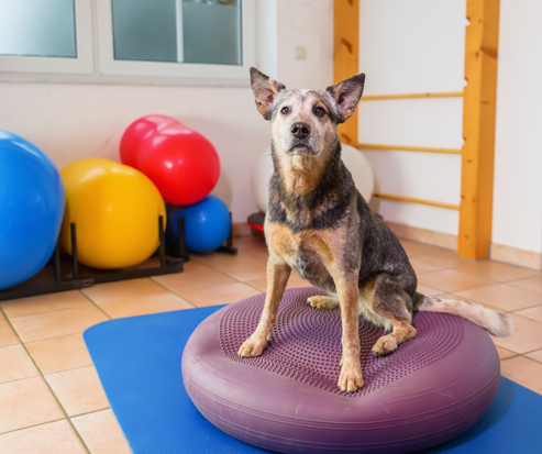
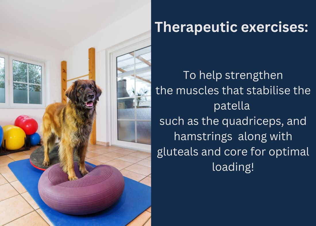
Balance and Proprioception Training Balance and proprioception exercises focus on improving the dog's awareness of limb placement and joint position, thereby enhancing stability and coordination. These exercises may include standing on unstable surfaces (e.g., balance pads or balance balls), walking over obstacles of varying heights, and performing controlled weight-shifting movements. Balance and proprioception training help retrain the neuromuscular system, reducing the risk of re-injury and improving overall functional performance. Hydrotherapy Hydrotherapy, which involves controlled exercises performed in water, plays a crucial role in the rehabilitation process for dogs recovering from knee injuries. Here's why hydrotherapy is considered an essential component of canine knee injury rehabilitation. Low-Impact Exercise Hydrotherapy provides a low-impact environment for dogs to exercise, allowing them to engage in physical activity without placing excessive stress on the injured knee joint. The buoyancy of water supports the dog's body weight, reducing the strain on the joints while still allowing for effective conditioning and muscle strengthening. Improved Range of Motion The resistance and buoyancy of water create a dynamic environment that promotes increased joint mobility and range of motion. Performing exercises in water allows dogs to move their joints through a greater range of motion than they might be able to achieve on land, facilitating joint flexibility and reducing stiffness. Muscle Strengthening Hydrotherapy exercises target and strengthen the muscles surrounding the knee joint, helping to stabilise the joint and prevent further injury. Resistance provided by the water encourages muscle engagement and development, promoting overall strength and endurance. Pain Relief and Reduced Inflammation The hydrostatic pressure of water can help reduce swelling and inflammation in the injured knee joint, providing natural pain relief. Immersion in warm water can also have a soothing effect on sore muscles and joints, promoting relaxation and alleviating discomfort. Cardiovascular Fitness Hydrotherapy sessions can be designed to incorporate aerobic exercises, such as swimming or walking on an underwater treadmill, to improve cardiovascular fitness. Aerobic conditioning helps increase stamina and endurance, enhancing the dog's overall fitness level and promoting weight management. Enhanced Proprioception and Balance Hydrotherapy exercises challenge the dog's proprioceptive abilities, or their sense of body awareness and position in space. Performing exercises in water requires dogs to adjust their movements to maintain balance and stability, thereby improving proprioception and coordination. Safe and Controlled Environment Hydrotherapy sessions are typically conducted under the supervision of trained professionals, ensuring that exercises are performed safely and effectively. Water depth, temperature, and resistance can be adjusted to meet the specific needs and abilities of each dog, making hydrotherapy suitable for dogs of all ages and fitness levels. Positive Reinforcement and Enjoyment Many dogs enjoy water-based activities, making hydrotherapy sessions a positive and enjoyable experience for them. The positive reinforcement associated with hydrotherapy can motivate dogs to actively participate in their rehabilitation program, leading to better compliance and outcomes. Nutrition and Monitoring Nutritional Support and Weight Management Proper nutrition and weight management are essential components of rehabilitation, especially for overweight or obese dogs. Maintaining a healthy body weight reduces stress on the joints and facilitates recovery from surgery. A balanced diet rich in high-quality protein, essential fatty acids, vitamins, and minerals supports tissue repair and muscle strength. Regular Monitoring and Adjustments Throughout the rehabilitation process, close monitoring by a veterinarian or certified rehabilitation therapist is essential. Progress should be assessed regularly, and the rehabilitation plan may need to be adjusted based on the dog's response to treatment, changes in condition, or any emerging issues. Open communication between the pet owner and the rehabilitation team is crucial to ensure the best possible outcomes for the dog's recovery. By incorporating these rehabilitation strategies and modalities into the post-operative care plan, pet owners can help their dogs recover from knee injuries more effectively, regain mobility, and improve their overall quality of life. It's important to work closely with veterinary and rehabilitation professionals to develop a tailored rehabilitation program that addresses the specific needs and capabilities of the individual dog. Canine knee injuries can significantly impact a dog's mobility and quality of life. Understanding the different types of knee injuries and the available pathways for rehabilitation is essential for effectively managing these conditions. Whether through conservative management or surgical intervention, coupled with appropriate rehabilitation protocols, we can help our furry friends regain function and live happier, more comfortable lives. If you want to learn more tricks and tips and be involved in a community of like-minded individuals, join our Facebook group at: The first time I ever heard about FHO surgery I was shocked to learn of the high success rates for doggo patients who have made the decision to undertake this somewhat radical approach to solving an all too common problem. As a hydrotherapist, planning a recovery program for a dog who has recently completed FHO surgery at first appeared daunting but when you have amazing patients and doggo owners who follow the plan step-by-step, what once appeared as daunting suddenly becomes one of the most rewarding rehabilitation cycles of my career. In this blog, we will dive into FHO surgery, what it entails and the plan towards a successful recovery using both hydrotherapy and physiotherapy as the main focal points on the journey to success.
What is FHO? Canine femoral head and neck excision (FHO) surgery is a procedure that addresses hip joint issues in dogs, providing relief and improved mobility. FHO is commonly performed to alleviate pain and discomfort associated with hip dysplasia, fractures, arthritis, or other conditions affecting the hip joint. It involves the removal of the femoral head and neck, allowing the formation of a "false joint" with surrounding muscles and tissues. During FHO surgery, the surgery removes the femoral head (the ball part of the ball-and-socket joint) and sometimes the neck of the femur. This eliminates the bone-on-bone contact that causes pain, and the body adapts by forming a pseudo-joint, utilising surrounding muscles for stability. The Road to Recovery Every journey starts out best with a proper plan or road map in place so that all parties involved are aware of the recovery process and what steps to take next to ensure the best recovery and rehabilitation possible. After FHO surgery, dogs will need rest and restricted activity. Pain medications and the beginning of some rehabilitation therapy will also be prescribed to manage discomfort and aid in the recovery process. Controlled exercise is crucial during the recovery phase. Short, on-leash/guided walks can help prevent muscle atrophy and promote joint mobility. Avoiding activities that place excessive strain on the hind legs, such as running or jumping is of the most importance at this early stage. Most Vets will ask patients to seek a physiotherapist post-surgery to aid and assist in the recovery process. Joint mobilisation exercises, massage and the use of laser therapy will all go a long way to promote strength and flexibility to aid in the recovery process as this is also a crucial time to be aware of potential complications that could quickly derail the road to recovery. These complications, such as infection or persistent lameness, need to be addressed as soon as they are noted and the safest option if there is ever any doubt would be to return to the vet for further review. As each patient progresses in recovery, the vet may gradually allow increased activity. However, it's essential to remain cautious and avoid pushing your dog too quickly, ensuring a steady and safe return to normal activities. This is the perfect time to add hydrotherapy to your recovery plan to not only continue in aiding the road to recovery but to also help build and manage muscles in the hind limbs and lower back. 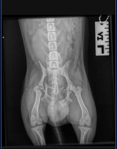
X ray taken pre-FHO
Hydrotherapy Case Study
If you want to learn more tricks and tips and be involved in a community of like minded individuals, join our facebook group at:
|
 RSS Feed
Physiotherapy joint mobilisation/ soft tissue massage
|
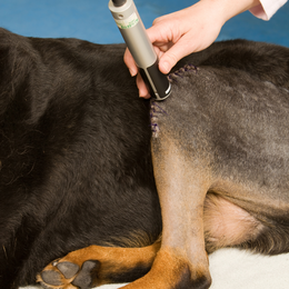
laser treatment
|
If your dog has been diagnosed with patella luxation, canine physiotherapy can be a very effective treatment option. By working with a qualified canine physiotherapist, you can help improve your dog's mobility, reduce their pain and discomfort, and improve their overall quality of life. So if you're looking for a non-invasive, holistic approach to treating patella luxation in your dog, consider canine physiotherapy at Canine Kinetics as an option.
Author
Joanna Whitehead ; Physiotherapist, Canine Physiotherapist & Hydrotherapist
Author
Joanna Whitehead
Matthew Ireland
Archives
June 2024
May 2024
April 2024
March 2024
February 2024
January 2024
December 2023
November 2023
October 2023
September 2023
August 2023
July 2023
June 2023
May 2023
March 2023
August 2022
April 2022
Categories
All
Canine Physiotherapist
Care And Guidance
Front Limb Injuries
Hydrotherapy
Myofascial Pain In Dogs
Neurological Conditions
Orthopaedic Condition
